The MICO Lab at USF is led by Dr. Margaret (Maggi) Mars Brisbin. The core focus of the lab is to unravel and interpret interactions between marine microbes in the context of changing ocean ecosystems. Overarching research aims are to determine how microbial interactions contribute to biogeochemical cycles and ecosystem function, how these interactions will respond to climate change, and how changes in the dynamics of these relationships will feedback on climate change impacts. The MICO lab uses multiple meta’omics and advanced microscopy techniques in conjunction with culture experiments and environmental sampling to tackle the following questions:
- What are the mechanisms of interaction between microbes in aquatic systems? How do phytoplankton or other hosts identify and select or recruit specific symbiotic partners? How are the relationships mediated? How does the relationship benefit the organisms involved, e.g., does the relationship provide a limiting nutrient or increase photosynthetic output?
- How do microbial interactions evolve? How quickly are new interactions established? How resilient are interactions to environmental change? Will climate impacts “break” or otherwise impact ecologically important microbial interactions?
- What are the ecological impacts if specific microbial interactions are disturbed? Will carbon fixation or sequestration decrease if interactions are diminished? How would such changes propagate through the food web?
The MICO lab is microbe agnostic and is generally intrigued by all interesting and ecologically relevant microbial interactions. Some systems of particular interest are acantharian-Phaeocystis symbioses, Phaeocystis colony-microbiome interactions, and bacterial interactions with the HAB-forming dinoflagellate Pyrodinium bahamense.
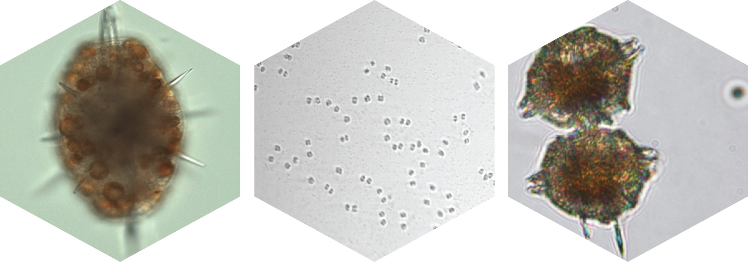
Left: An acanatharian with Phaeocystis antarctica endosymbionts collected from the Southern Ocean on the Palmer LTER cruise and imaged with Prakash Lab custom microscopes; Center: A Phaeocystis antarctica colony speckled with bacteria that was imaged with the Imaging Flow Cytobot aboard the R/V Nathaniel B. Palmer in the Southern Ocean; Right: Pyrodinium bahamense cells collected in Old Tampa Bay and imaged with light microscopy by FWRI.